Character description
-
- species
To enter the species name, if available.
- genus
To enter the name of the species’ genus, if available.
- family
To enter the name of the species’ family, if available.
- ID information
Genebank number / collection ID
- collector
To enter the name of the person who collected the specimen.
- country
The country in which the specimen/lot was collected.
- locality 1
The political subunit (e.g., departamento/state) in which the specimen/lot was collected.
- locality 2
The second smaller political subunit (e.g., provincia/county) in which the specimen/lot was collected
- coordinates 1
The longitude parameters of the coordinates where the specimen/lot was found, given as decimal value.
- coordinates 2
The latitude parameters of the coordinates where the specimen/lot was found, given as decimal value.
- Gosner stage
Staging of the specimen after Gosner (1960). In cases of heterogeneous lots each diverging specimen has to be entered as a new data set. Only specimens between GS 32-40 are considered.
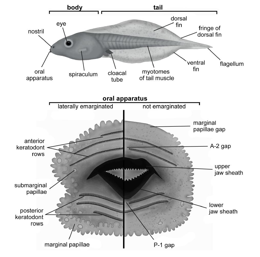
- total length
The total length (mm) from head to tail tip of the tadpole, presented with to decimal places.
- body length
The body length (mm) of the tadpole, measured laterally from tip of snout to the junction of the ventral edge of the tail muscle with the body, presented with two decimal places.
- tail length
The tail length (mm) of the tadpole, calculated from total length – body length.
- maximum body height
The maximum body height (mm) measured in lateral view.
- maximum body width
The maximum width (mm) of the body measured in dorsal view.
- snout-nostril distance
The distance (mm) measured from the foremost tip of the snout to the centre of the nostrils in lateral or dorsal view.
- snout-spiraculum distance
The distance (mm) measured from the foremost tip of the snout to the centre of the spiraculum opening in lateral or ventral view.
- eye diameter
The diameter of the eye (mm) measured in lateral view from the most left to the most right corner of the eye.
- internostril distance
The distance (mm) measured between the centres of the nostrils in dorsal view.
- interorbital distance
The distance (mm) measured between the centres of the eyes at level of iris in dorsal view.
- maximum tail height
The height of the tail in lateral view measured at the highest point of the tail including the ventral and dorsal fins.
- maximum height of dorsal fin
The maximum height of the dorsal fin measured laterally from the upper border to tail muscle at point of maximal tail height.
- maximum height of ventral fin
The maximum height of the ventral fin measured from the lower border to tail muscle at point of maximal tail height.
- maximum tail muscle height
The height of the tail muscle in lateral view measured at the highest point of the muscle near or at the tail muscle base (where the ventral line of the musculature meets the contour of the trunk) excluding the ventral and dorsal fins.
- tail muscle width
The width of the tail muscle in dorsal view measured at the widest point of the muscle near or at the tail muscle base.
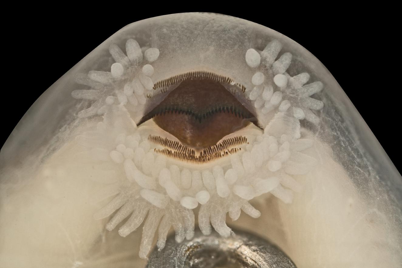
- oral disc width
The width (mm) of the oral disc in ventral view in (usual) semi-opened position.
- position of oral disc
The position of the disc in lateral view: ventral, near-ventral, anteroventral, almost-anterior, anterior, anterodorsal, dorsal.
- disc emarginated
To enter information about a laterally, anteriorly and/or posteriorly emarginated oral disc.
- LKRF
To enter the entire keratodont row formula beginning with the number of the anterior rows (counting from the tip of the snout) followed by rows with gaps in parentheses; jaws indicated by “/”; followed by all posterior keratodont rows with those having a gap in parentheses: e.g.; 3(1)/3(3); rows having gaps only in some specimens are given in squared brackets: 3(1)[2]/3(3)[1]; superscript numbers indicate bi-or triseriate keratodont rows (e.g., in Ascaphus, Alytes); no keratodont rows but jaw sheaths are present “0/0”, if no structures are present, “absent”, and if structures are obviously incomplete “destroyed” has to be noticed.
- anterior keratodont rows
The number of all anterior keratodont rows.
- posterior keratodont rows
The number of all posterior keratodont rows.
- anterior gaps
To enter yes/no if anterior gaps are present.
- anterior rows with gaps
The number of anterior rows with gaps in total (not the specific row with the gap).
- posterior gaps
To enter yes/no if posterior gaps are present.
- posterior rows with gaps
The number of posterior rows with gaps.
- rel. length ant. rows
To indicate the proportional length of al anterior rows: e.g., A-3<2=4<1 meaning that A-3 is the largest row, followed by rows A2 and A4 of the same length but still longer than A-1 row; length of rows considered in relation to the entire oral disc but not including their gaps so that a row with a very wide gap and thus two short fragments is still treated as being longer than a neighbouring row without gaps but with shorter ends.
- rel. length post. rows
To indicate the proportional length of al anterior rows: e.g., P-1=2<3 (see explanation of no. 73).
- anterior papillae
To indicate the rows of marginal papillae in the region of the anterior labium, alternating rows indicated by “b”, eg., 2b means two alternating anterior marginal papillae rows, “2-3” if in this region 2 to 3 rows are present, or “absent” if no papillae are present, or “irregular” if papillae are presented irregularly spread all over the outer margin and the inner membrane of the oral disc (and no differentiation in marginal or submarginal papillae is possible).
- posterior papillae
To indicate the rows of posterior marginal papillae, alternating rows indicated by “b”, eg., 3b means three alternating posterior marginal papillae rows, “2-3” if in this region 2 to 3 rows are present, or “absent” if no papillae are present, or “irregular” if papillae are presented irregularly spread all over the outer margin and the inner membrane of the oral disc (and no differentiation in marginal or submarginal papillae is possible).
- MPRF
The marginal papillae row formula indicates the number of rows (e.g., 1=uniseriate, 2=biseriate, et cetera), gaps in those rows in parentheses, a hyphen indicating a change in the number of rows of marginal papillae in the respective region of the oral disc, and a “b” indicating alternating rows; e.g., (1)/2b/2b-3 = marginal papillae uniseriate anteriorly with (wide) gap, alternating biseriate laterally, and alternating biseriate to triseriate posteriorly.
- shape of marginal papillae
Describing the shape of the papillae as either (or both) triangular, conical (blunt -ended with triangular shape), blunt (without any tapering), tubercle-like (short), round (tubercle-like), enlarged (barble-like).
- submarginal papillae
To state whether submarginal papillae are present “yes”, “no”, or “irregular” if papillae are presented irregularly spread all over the outer margin and the inner membrane of the oral disc (and no differentiation in marginal or submarginal papillae is possible).
- jaw sheaths present
To state whether jaw sheaths are present (yes) or not (no).
- jaw sheaths serrated
To state whether the jaw sheaths are slightly, finely, clearly, heavily, or no, if not serrated.
- upper jaw sheath shape
To enter the shape of the upper jaw sheath: moderately arched, broadly arched, narrowly arched, saddle, m-formed, triangular, except its ends unbowed, divided bars, or half-divided bar).
- lower jaw sheath shape
To enter the shape of the lower jaw sheath: v, broadly v, slightly v, u, broadly u, divided, M-formed with two peaks, or curved down.
- jaw sheaths thickness
Information about the thickness of the jaw sheaths (especially the upper one) in regard to their length (modified after Anstis 2014): stated as slender (if jaw sheaths have less than half the depth of the upper/lower jaw cartilage keratinised and the sheaths appear thin compared to the entire oral disc), medium (1/2 to ¾ of the cartilages moderately keratinised), robust (cartilages fully or almost keratinised with jaw sheaths appearing prominent), massive (cartilages extremely heavily keratinised with jaw sheaths appearing very thick), or two characters if upper and lower jaw sheath are of different thickness (e.g., slender/massive).
- nostrils present
To enter yes/no whether openings of the nostrils in the epidermis are present or not.
- position of nostrils
The position of the nostrils: dorsally, dorsolaterally, anterodorsally, laterally, anterolaterally or anteriorly.
- direction of nostrils
The direction in which the nostrils are orientated: dorsally, dorsolaterally, laterally, anterolaterally or anteriorly
- shape of nostrils
To enter the shape of the nostrils: ovoid, drop-shaped, reniform, angularly, slotted, or round.
- nostrils with structure
To indicate whether an entire operculum (eo), or half (or less covering) operculum (ho), a rounded bulge or torus, a half torus, a sulcus between nostrils and eye, notched tube, one or more papillae-like structures around the opening, or no operculum or other structure is present.
- position of eyes
The position of the eyes: dorsally, dorsolaterally or laterally.
- direction of eyes
The direction in which the eyes are orientated: dorsally, dorsolaterally, laterally, lateroventrally, or anteriorly.
- position and size of spiracle
The position and the size of the spiracle (not only of its opening), orientated from the median axis of the lateral tadpole: low-sinistral-short (lss), low-sinsitral-long (lsl), low-sinistral-just-opening (lso), median-sinistral-short (mss), median- sinistral -long (msl), almost-ventral-sinistral-short (vss), median (single or fused, next to vent tube)-posterior-ventral (spv), single-midventral in posterior third of the body (mpv), single-midventral in anterior third of the body (mav), single ventrolaterally in posterior third of the body (svl), posterior-sinistral (median) short (pss), posterior-sinistral (median) long (psl), median-sinistral in posterior third of the body (msp).
- shape of spiracle
The shape of the spiracle: inner (=centripetal) wall absent, inner wall present as slight ridge, inner wall up to ¾ free from body, spiracle opens laterally (instead of posteriorly), entire (lateral or medial-posterior) tube, medial broad, flabellate tube, or median slit.
- position of vent tube (opening)
The position of the vent orientated by the direction of its aperture: medial a: medial vent tube opening medially, medial b: medial vent tube with dextral displacement or dextrally directed opening, dextral a: opens partway or midway on the right side up from the lower margin edge of the ventral fin, dextral b: opens down to right side margin of the ventral fin, dextral c: opens beneath a long skin fold with the vent tube shorter than this fold (on the right side of the ventral fin), dextral d: opens (as a slit rather than a tube) on the right side of the ventral fin in ventral view beneath a broad pouch of skin which covers the hindlimb buds until they first emerge at higher developmental stages, dextral e: opens on the right side of the ventral fin directly below tail muscle and (at least partly), covered by the hind limb, dextral f: opens on the right side of the ventral fin, ventrally directed, not attached to ventral fin and covered by hind limb, dextral g: opens on the right side at the ventral margin in a dextral-ventral direction, fully attached to the fin in a bulgy fold, dextral h: opens on the right side at ventral margin, entirely covered by median anal flap, dextral i: opens at the right margin of the ventral fin as a remarkable elongated tube.
- shape of vent tube (in lateral view of right side of supine tadpole from head to tail)
The shape of the vent tube (displaced = wall shorter in that region/direction): right wall displaced dorsally, right wall displaced anteriorly, right wall displaced anteriorly and dorsally, right wall displaced posteriorly and dorsally, right wall displaced posteriorly, dextral tube with equal walls, dextral opening not formed as a tube, medial vent tube with equal walls, medial vent tube with ventral wall displaced posteriorly, medial vent tube with ventral wall displaced anteriorly.
- attachment of vent tube to ventral fin
The attachment of the vent tube to the dorsal fin: vent tube with web between tube and fin (a), vent tube fully attached to ventral fin (b), or not attached (c).
- lat. line system visible
To enter yes/no whether neuromasts of a lateral line system are visible or not.
- position of lat. line system
Describing the position/run if neuromasts are visible forming a line: dorsally, dorsolaterally, laterally (multiple entries possible if two or more “lines”), or absent if not present (referring to cell 99).
- rel. height of fins
To describe the height of fins correlated to each other: (d “=/” v; “>=”to indicate if the dorsal fin is only barely higher than the ventral fin)
- dorsal fin reaches onto body
To enter yes/no whether the dorsal fin emerges before the body-tail junction or not.
- margin and origin of dorsal fin (rising)
Describing the shape of the dorsal fin: dorsal fin begins at the body-tail junction with a (shallow) convex raising (a), dorsal fin begins up to one-third onto the body with a moderate-convex rising (b), dorsal fin well arched beginning midway or more along the body (c), dorsal fin abruptly arched anteriorly with a convex margin (d), dorsal fin moderately arched posteriorly with the rising well posteriorly of the body-tail junction (e), dorsal fin rises on body slightly before body-tail junction in moderate convex rising (f), dorsal fin nearly without any rising and with horizontal shape (g).
- margin of ventral fin
Describing the shape of the ventral fin: ventral fin with a shallow (nearly) horizontal margin (a), ventral fin convex, deeper than dorsal fin (b), ventral fin (well) convex arched (c), ventral fin (moderately) arched posteriorly with the rising well posteriorly of the body-tail junction (d), ventral fin triangular shaped (usually deeper than dorsal fin) and tapered (e).
- tip of tail
Information about e.g., a broad triangular, finely acuminate, round, pointed, tail tip or ending in an acuminate flagellum, elongate flagellum, or a short flagellum.
-