Global Tadpole Database

Global Tadpole Database
| Home | Search Database | Log-In | Impressum |
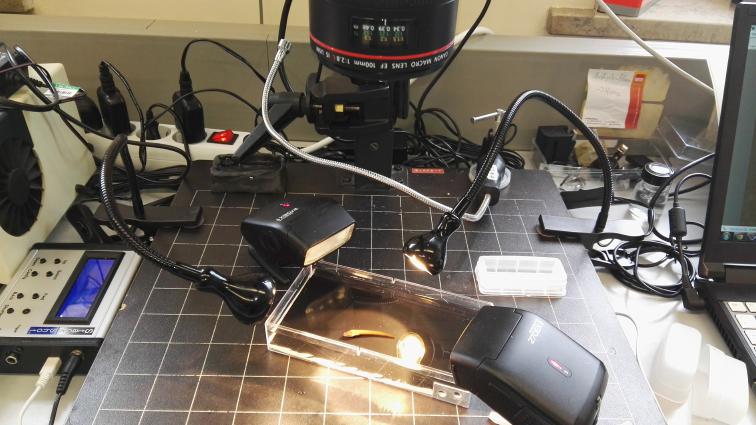
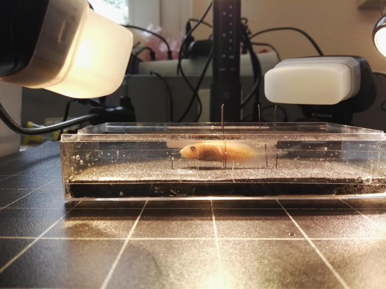
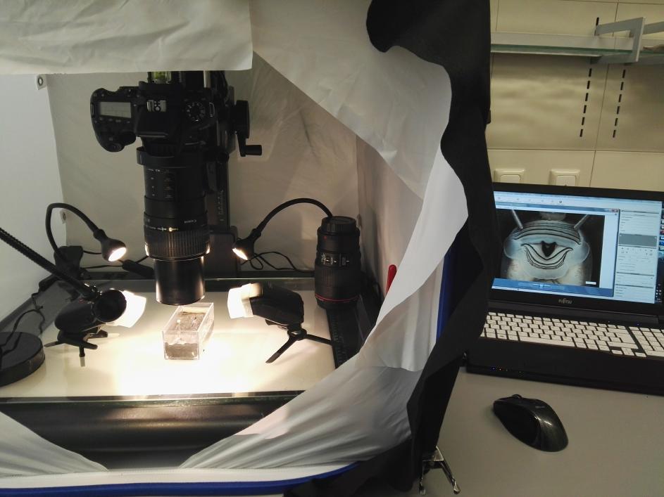
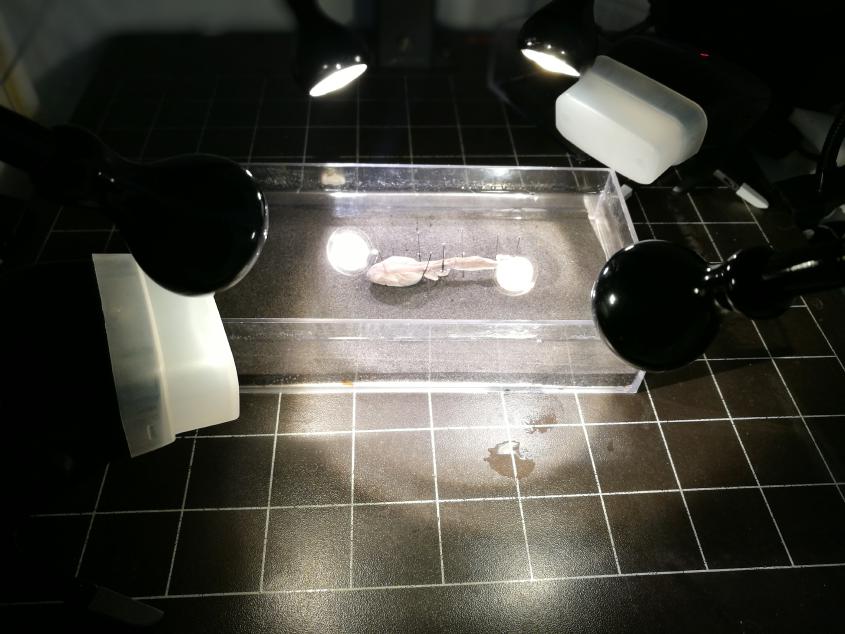
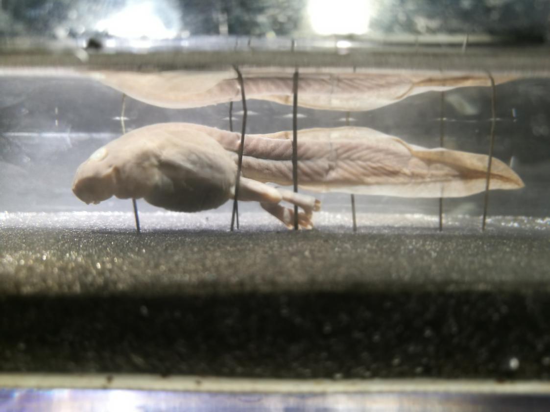
A setup of a SLR camera and magnifier lenses (Canon 100 mm 2.8L and Canon MP-E 65 mm) mounted on an electronic-driven macro rail was used for the digital images. A stack of 10-15 images was taken and merged using Helicon Pro software to achieve images with a high depth of focus. Morphological descriptions and measurements were done on the basis of digital and scaled images of preserved tadpoles. Terminology of morphological characters follows Altig and McDiarmid (1999). To identify developmental stages Gosner’s (1960) classification was used. Structures of the oral apparatus were described according to Altig (1970), except for the term “keratodont”, which is used for the keratinized structures on the labia of the oral disc. Marginal papillae are considered separately for the region of the upper labium, the lateral region, and the region of the lower labium and the “marginal papillae row formula” (MPRF) is provided according to Schulze et al. (2015).